PET, PET/CT, MicroPET, PET/MRI
Positron Emission Tomography (PET)
Positron emission tomography (PET) provides an ideal technique of in-vivo, non-invasive functional imaging in subjects. It is a nuclear medical imaging technique that produces three-dimensional image of functional processes in the body. A small amount of labelled compound (radio-pharmaceutical or radiotracer) is introduced into the subject and after an appropriate uptake period, the scanner measures the concentration of tracer in tissue. Three-dimensional images of tracer concentration within the body are constructed by computer analysis. Subjects are imaged under general anaesthesia.
At the Department of Clinical Medicine a High Resolution Research Tomograph (HRRT) Scanner from CPS Innovations is available at the PET centre´s research division, building 10, Nørrebrogade 44, 8000 Aarhus C. The expertise at the PET centre´s research division primarily focuses on pigs, however it is in principle possible to PET scan all species of animals from rodents to human size in the scanner. While scanning it is possible to sample series of arterial blood for the establishment of the time-activity curves (CAT), which is used for kinetic modeling of the tracer substance distribution. Blood samples can also be used for metabolite analysis.
The centre has available all equipment for the scanning procedure both surgical and for monitoring the anaesthetized subject. Further and high-resolution analysis can be performed of tissue sections of identified areas of interest by autoradiography (Fujifilm IP Eraser Bio-Imaging Analyzer), which allows for high resolution of a tissue section.
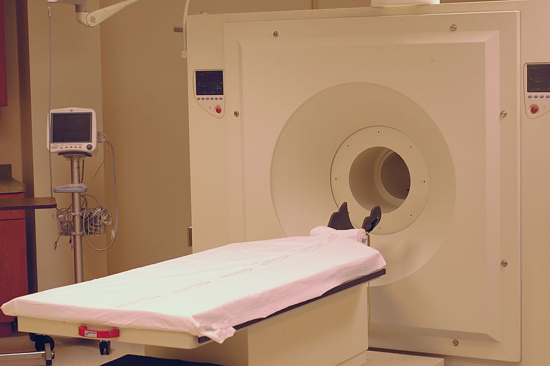
PET/CT
Computed Tomography (CT) refers to imaging by sections or sectioning, through the use of any kind of penetrating wave. CT is a technology that uses computer-processed x-rays to produce tomographic images (virtual 'slices') of specific areas of the scanned subject. This allows the investigator to see what is inside it without cutting it open. A PET/CT scanner from Siemens is available for animals at the PET centre´s research division. It combines PET functional imaging with CT tomographic imaging and detailed three-dimensional anatomical images are obtained. This is very useful in showing detailed views of moving organs or structures with higher anatomical variation. PET/CT images are used for diagnostic and therapeutic purposes in various medical disciplines.
The PET/CT scanner is located at the PET centre´s research division, building 10, Nørrebrogade 44, 8000 Aarhus C. Though the expertise in the PET centre´s research division is primarily focused on pigs it is possible to perform PET/CT scans on species the size of rabbits up to human size in the scanner. The resolution is too poor for small animals like rodents. As for PET it is possible to sample series of arterial blood for the establishment of the time-activity curves (CAT) while scanning. Samples are used for kinetic modeling of the tracer substance distribution. Blood samples can also be used for metabolite analysis.
MicroPET
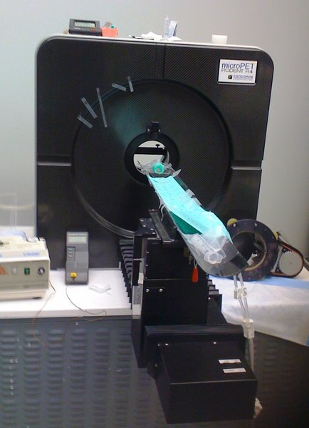
Is a high-resolution positron emission tomography (PET) technology designed for imaging rodent or small laboratory animals. A microPET Rodent R4 from Concorde Microsystems, Inc. is available at the PET centre. Two MicroPET scanners are available at the centre. As for PET and PET/CT it is possible to sample series of arterial blood for the establishment of the time-activity curves (CAT) while scanning. Samples are used for kinetic modeling of the tracer substance distribution. Blood samples can also be used for metabolite analysis.
PET/MRI
Is a hybrid imaging technology that combines MRI soft tissue morphological imaging and PET functional imaging. At the PET centre´s research division a nanoScan®PM PET/MRI system for small animal imaging from Mediso Medical Imaging Systems is available. This compact imaging system utilises magnetically shielded position sensitive photomultiplier tubes and a compact 1 Tesla permanent magnet MRI platform. This provides high spatial resolution of down to 100 µm, with excellent soft tissue contrast required for anatomical/morphological imaging.
Equipment, facilities and price
Equipment
The scanning equipment described above is located at the Department of Clinical Medicine at the PET centre´s research division, building 10, Nørrebrogade 44, 8000 Aarhus C. All surgical equipment and equipment to monitor the anaesthetised subject during the scanning procedure are available at the PET centre.
Please note that scanning equipment cannot be moved from this location.
Facilities
For housing of experimental animals (rodents) there are two animal facilities associated with the PET centre.
Price
A cost is associated with usage of the imaging technologies described above. Please inquire for price with Aage Kristian Olsen Alstrup (see above).
Contact
Aage Kristian Olsen Alstrup (e-mail aagols@rm.dk) who will be able to provide detailed information concerning the use of equipment and method as well as the guidance on the necessary requirements for experimental setup (application for experimental licence, capabilities and use of the equipment etc.).